Surgery109
Recurrence of diaphragmatic hernia in a 21-year-old man - case report.
Nawrót przepukliny przeponowej u 21 letniego mężczyzny – opis przypadku.
Marcin Neska✉1Dominik Glinka1Małgorzata Neska-Matuszewska2Michał Matuszewski3
1Uniwersytet Medyczny im. Piastów Śląskich we Wrocławiu
2Katedra i Zakład Radiologii Ogólnej, Zabiegowej i Neuroradiologii, Uniwersytet Medyczny im. Piastów Śląskich we Wrocławiu
3Katedra i Klinika Urologii i Onkologii Urologicznej, Uniwersytet Medyczny im. Piastów Śląskich we Wrocławiu
✉Marcin Neska
Otrzymano: 11.07.2018Zaakceptowano: 04.12.2018Opublikowano: 06 December 2018
e-ISSN: 2084-2708 (primary version)p-ISSN: 2083-0033 (print version)
Streszczenie
Wstęp
Wrodzona przepuklina przeponowa (WPP) jest wadą rozwojową polegającą na przemieszczeniu narządów jamy brzusznej w obręb klatki piersiowej. Przemieszczenie następuje przez otwór w przeponie powstały w okresie płodowym. Do nawrotu przepukliny przeponowej dochodzi rzadko, najczęściej po kilku tygodniach od operacji.
Opis przypadku
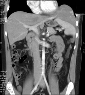 21-letni mężczyzna zgłosił się z nawracającymi dolegliwościami brzusznymi, nasilającymi się po spożyciu posiłku. W 4 dobie życia chory operowany z powodu WPP, w wieku 8 lat przeszedł zabieg appendektomii. Kilkukrotnie występowała u chorego niedrożność przewodu pokarmowego, poddająca się leczeniu zachowawczemu. USG jamy brzusznej wykazało strukturę o echogeniczności wątroby zlokalizowaną atypowo – nad śledzioną, bez cech hepatomegalii. Następnie, tomografia komputerowa (TK) jamy brzusznej z kontrastem dożylnym wykazała przemieszczenie lewego płata oraz segmentu IV wątroby do klatki piersiowej nad lewą kopułą przepony.
Wnioski
U osób po rekonstrukcji WPP zgłaszających dolegliwości gastryczne należy podejrzewać nawrót przepukliny przepony. U opisywanego chorego nawrót WPP nastąpił najprawdopodobniej w okresie noworodkowym i przez wiele lat nie został rozpoznany.
Keywords:przepuklina przeponowaból brzuchanawrót przepukliny przeponowej
Abstract
Introduction
Congenital diaphragmatic hernia (CDH) is a developmental defect consisting in the dislocation of abdominal organs into the chest. The displacement occurs through a hole in the diaphragm formed during the fetal period. The recurrence of diaphragmatic hernia is rare and appeared most often after a few weeks after surgery.
Case Report
A 21-year-old man reported for recurrent abdominal pain, impairing after eating a meal. The patient was operated due to CDH on the 4th day of life and at the age of eight he underwent appendectomy. Several times the patient had a gastrointestinal obstruction, undergoing conservative treatment. The abdominal ultrasound showed a structure with liver echogenicity localized atypically – above the spleen, with no sign of hepatomegaly. Subsequently, Computer Tomography (CT) of the abdominal cavity with intravenous contrast showed the presence of the left lobe and segment IV of the liver in the chest above the left dome of the diaphragm.
Conclusions
People who have reconstructed the CDH and reporting gastric complaints should be suspected for diaphragmatic hernia recurrence. It is possible that CDH recurrence appeared shortly after surgery in the neonatal period but it was not recognized for many years.
Keywords:diaphragmatic herniaabdominal paindiaphragmatic hernia recurrence
References
- 1.Czernik J. Chirurgia dziecięca. 2005. PZWL
- 2.Dobrzańska A., Ryżko J. Pediatria. Podręcznik do Państwowego Egzaminu Lekarskiego i egzaminu specjalizacyjnego. 2010. Urban&Partner
- 3.Wright J.C., Budd J.L., Field D.J., Draper E.S. Epidemiology and outcome of congenital diaphragmatic hernia: a 9-year experience. Paediatric and Perinatal Epidemiology. 2011:144-149
- 4.4. de Buys Roessingh A.S., Dinh-Xuan A.T. Congenital diaphragmatic hernia: current status and review of the literature. Eur J Pediatr. 2009:393-406
- 5.5. Conforti A.F., Losty P.D. Perinatal management of congenital diaphragmatic hernia. Early Hum Dev. 2006:283-287
- 6.6. Elhalaby E.A., Abo Sikeena M.H. Delayed presentation of congenital diaphragmatic hernia. Pediatr Surg Int. 2002:480-485
- 7.7. Brindle M.E., Brar M., Skarsgard E.D. Canadian Pediatric Surgery Network (CAPSNet). Patch repair is an independent predictor of morbidity and mortality in congenital diaphragmatic hernia. Pediatr Surg Int. 2011:969–974
- 8.8. Snoek K.G., Reiss I.K.M., Greenough A. et al. Standardized postnatal management of infants with congenital diaphragmatic hernia in Europe: the CDH EURO consortium consensus—2015 update. Neonatology. 2016:66–74
- 9.9. Moyer V., Moya F., Tibboel R. et al. Late versus early surgical correction for congenital diaphragmatic hernia in newborn infants. Cochrane Database Syst Rev. 2002
- 10.10. Wung J.T., Sahni R., Moffitt S.T. et al. Congenital diaphragmatic hernia: survival treated with very delayed surgery, spontaneous respiration, and no chest tube. J Pediatr Surg. 1995:406–409
- 11.11. Tsao K., Lally P.A., Lally K.P. Congenital Diaphragmatic Hernia Study Group. Minimally invasive repair of congenital diaphragmatic hernia. Journal of pediatric surgery. 2011:1158-1164
- 12.12. Putnam L.R., Tsao K., Lally K.P., et al. Minimally-Invasive vs Open Congenital Diaphragmatic Hernia Repair: Is There a Superior Approach?. Journal of the American College of Surgeons. 2017:416-422
- 13.13. Hajer G.F., vd Staak F.H., de Haan A.F., Festen C. Recurrent congenital diaphragmatic hernia: which factors are involved?. Eur J Pediatr Surg. 1998:329–333
- 14.14. Grethel E.J., Cortes R.A., Wagner A.J. et al. Prosthetic patches for congenital diaphragmatic hernia repair: Surgisis vs Gore-Tex. J Pediatr Surg. 2006:29-33
- 15.15. Fine R., Borrero E., Stone A. Bochdalek hernia in adulthood. N Y State J Med. 1987:516-518
- 16.16. Hines G.L., Romero C. Congenital diaphragmatic hernia in the adult. Int Surg. 1983:349–351
- 17.17. Nagata K., Usui N., Terui K., et al. Risk factors for the recurrence of the congenital diaphragmatic hernia-report from the long-term follow-up study of Japanese CDH study group. Eur J Pediatr Surg. 2015:9-14
- 18.18. Sato K., Orihashi K., Hamanaka Y. et al. Post-traumatic diaphragmatic herniation of the liver, examined by positron emission tomography: case report. World Journal of Emergency Surgery. 2011:30