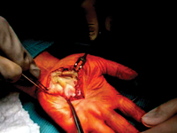
Isolated hand plexus neurofibroma: two case reports. Diagnostics and treatment proposals.
Neurofibromas
Plexiform neurofibromas are benign tumors of the peripheral nerve sheath. They can be found as single or multiple nodules, which are part of the
of the clinical picture in any type of neurofibromatosis, and can become malignant. Neurofibromas arise from Schwann cells
which are part of the nerves of the skin throughout the body, nerves innervating internal organs and cranial nerves. The most characteristic symptoms include the presence of
scattered, elongated, resilient subcutaneous nodules. These are fibromas,
histologically resembling individual neurofibromas, appearing along the
the course of peripheral nerves or cranial nerves. They most often occur on the
face and neck. Sometimes they lead to massive enlargement of a limb or other
part of the body (neurofibromatous elephantiasis) [1,2]. Another equally common
common symptom is skin discoloration, looking
resembling coffee spots ("giant café-au-lait spot"),
as well as freckle-like lesions localized around the
axillae and axillary pits [3]. Neurological symptoms are associated
with the compression of neurofibromas on adjacent structures. The patient
then complains of neuralgia of the peripheral nerves. In the literature we can find isolated
cases of neurofibroma plexiformis without any association with neurofibromatosis
type 1 [4-6]. Treatment
of patients with plexiform neurofibromas is not well defined
defined and is mainly aimed at controlling symptoms.
Surgical excision is the only available therapy, as there
there are no drugs that can prevent their formation or treat them conservatively
conservatively. At present, the results of surgical excision
are not fully satisfactory, and these procedures can be complicated
due to the size, location, condition of the vessels, involvement of the
neural involvement, microscopic staging and high rates of tumor re-growth
[7]. It should
also take into account the fact that there are known
there are many causes causing tumor-like lesions
to neurofibromas .
These include traumatic soft tissue injuries,
arthritis,
tendon diseases (e.g. tendinosis, tendon sheath inflammation,
tendon rupture), non-neoplastic soft tissue lesions (e.g.
ganglion, bursitis, granuloma)
,
and, less commonly, tumors and their metastases to soft tissues [8].
Focal lesions of soft tissues
soft tissues are often the cause of pain and
palpable tumor mass
[9].
W
these cases, especially in the early stages, accurate
diagnosis
is crucial,
to establish a diagnosis and treatment strategy. Therefore,
w
when confronted with a focal tumor lesion
the logical
seems
is to follow
established algorithm of
procedure. First, it should provide an accurate
description
of the lesion in correlation with the patient's age, location, type of involved
tissue,
the condition of the adjacent
environment.
For this purpose, in addition to
detailed clinical examination, ultrasound should be performed
of the occupied
area, and
if a bony lesion is suspected then an x-ray examination in two projections with adjacent joints. The advancement of
bone lesions, it is necessary to
deepening
diagnosis through CT examination.
However,
lesion
limited
only to soft tissues on ultrasound will require
MRI diagnosis,
which
will more accurately visualize the infiltration
by
its neighborhood.
If imaging studies show a high
probability of proliferative disease,
an important diagnostic element is to obtain material for
pathomorphologic examination, taken during open biopsy or
oligobiopsy and establish the histological diagnosis [10].
If the neoplastic process is confirmed, it is always necessary to
X-ray/CT scan of the chest, ultrasound/CT scan of the abdomen and even
even PET to exclude metastases [11]. The second
stage of the implemented algorithm dictates that treatment should be planned based on the
answers to the following questions: - Is the lesion likely to be a
normal variant, i.e., "leave me alone, don't touch me."
lesion of a benign type, requiring no further imaging or
supplies, but only observation? [12]
- Does the lesion have features suggestive of an aggressive nature, and then how
far advanced
should
be
treatment? [13].
The heterogeneity of the clinical picture of unidentified focal
nodular lesions in the metacarpal region,
the variety of available methods of diagnosis do not make the task any easier
the task, both at the stage of making the initial diagnosis and
during communication between members of the interdisciplinary team,
who, on more than one occasion, should handle the treatment. Therefore, it seems important to cooperate on the line of
orthopedist-oncologist-pathomorphologist. Two cases of isolated neurofibromatosis of the palm of the hand
are described below because of their uniqueness. The second of the described cases indicated an additional possibility of postoperative treatment of the patient in the form of prolotherapy injections to improve the function of the treated limb.