News
131 entries in total
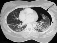
Recurrent pneumothorax in course of cystic fibrosis – treatment challanges. Case report.
Cystic fibrosis (cystic fibrosis) is the most common genetic disease with autosomal recessive inheritance among the Caucasian population. The incidence varies from country to country. In Poland, it is about 1/2300 of live births. In its course, as a result of mutations on both copies of the CFTR (cystic fibrosis transmembrane conductance regulator) gene, which encodes a chloride channel, ion transport in all exocrine glands is disrupted, leading to dysfunction of key organs for survival, such as the pancreas, liver, lungs and intestines [1].Respiratory complications are the first cause of premature death in CF patients. Pulmonary manifestations of CF include hemoptysis, pneumothorax, chronic sinusitis and airway inflammation, bronchial dilatation, and respiratory failure, among others. Pneumothorax is the presence of air in the pleural cavity. Risk factors are associated with chronic infection with Burkholderia cepacia and Pseudomonas aeruginosa and the coexistence of other complications such as hemoptysis and pancreatic insufficiency [4]. Significant disease progression (FEV1 30%) [2] also favors the occurrence of this complication. Taking into account the criterion of time, we can distinguish the first case of emphysema, or recurrent emphysema, when the air leak reappears within 7 or more days after the previous episode. If the air leak persists for 5 or more days, we can recognize persistent emphysema (Shidlow 1993). Therapy can be divided into medical (oxygen therapy and observation) or surgical interventions. The latter group, which includes drainage, pleurodesis and pleurectomy depending on the course of treatment, is considered a more effective therapeutic option for patients with emphysema without significant concomitant lung disease. However, in CF-affected patients, it may complicate the lung transplant procedure in the future. There are no clinical trials available that reliably compare the efficacy and risk of complications of both types of interventions in CF patients [3-4]. An additional complication is that emphysema recurs in 50-90% of these patients, and in almost half of them it also occurs on the opposite side of the chest. Emphysema significantly worsens lung function, and the patient experiences shortness of breath and significant pain. If refractory to treatment, it is an independent indication for lung transplantation.[5]Here we present the case of a 16-year-old female patient with recurrent unilateral pneumothorax, in which, due to the severity of the disease, the diagnosis and therapeutic measures taken were fraught with numerous problems.
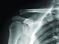
Migration of a Kirschner wire used in fixation of acromioclavicular joint dislocation – case report.
Dislocation of the acromioclavicular joint refers to complete or partial misalignment of the articular surfaces of the neighbouring scapula and clavicle [1]. It is an injury which often occurs in contact sports, such as football, judo, rugby and many more. It is also a relatively common injury in daily activities, as a result of falling during a walk or falling off a chair with impact on the shoulder girdle [2], [3].<br />Depending on the grade of the injury, recommended approaches vary from conservative treatment (grade I and II) to surgical treatment (above grade III).<br />Conservative treatment recommendations are: cooling, painkillers and limb elevation. Surgical treatment consists of surgical stabilisation of the dislocation. There are many techniques and methods to fixate the injury, including: Kirschner wires, using TightRope, double Endobutton, MINAR, Copeland and Kessel, Waver and Dunn or dedicated plates [3], [4].<br />
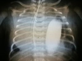
Recurrence of diaphragmatic hernia in a 21-year-old man - case report.
Congenital diaphragmaƟc hernia (CDH) is a developmental defect consisƟng in the dislocaƟon of abdominal organs into the chest. The displacement occurs through a hole in the diaphragm formed during the fetal period. The recurrence of diaphragmaƟc hernia is rare and appeared most oŌen aŌer a few weeks aŌer surgery.
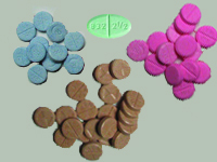
Effect of vegetarian diet on INR in patient treated with anticoagulants
<p>Warfarin sodium (Latin: Warfarinum natricum), found under the trade name Warfin, is an effective anticoagulant used in the prevention and treatment of thrombosis. It is widely used after cardiac surgery, especially after implantation of an artificial heart valve prosthesis. Warfin's role is to prevent blood from clotting in the prosthesis and valve components and to help dissolve any clots that form.</p><p>Patients must continue anticoagulant treatment for the rest of their lives. They also remain under the constant medical care of the cardiology clinic. In general, they should monitor the prothrombin time (PT), or the time of blood clot formation, every 4-6 weeks to see how the clotting factors that go into the so-called prothrombin syndrome are working. The result is presented under the name International Normalized Ratio (INR) (<i>Enternational</i><i>Normalized Ratio)</i> [1]. In order to maintain stable blood density values, patients should adhere to a well-balanced diet, thus ensuring that the body receives the recommended daily dose of vitamin K contained in foods. Too high a dose of vitamin K in relation to the body's needs can significantly affect the INR value in the blood tested and create difficulties in selecting the antivitamin K dose [2].</p><p></p><p>There are three classified forms of vitamin K: K1 and K2 - derived from a natural source, and K3 which is a synthetic chemical compound. Vitamin K1 (phylloquinone) is synthesized exclusively in plants and taken with food, while K2 is a group of compounds known as menaquinones (MK-n) [3]. These compounds are synthesized by intestinal bacteria. They are mainly found in animal products, including those that are fermentable (Table 1).</p><p>Table 1.</p><p>Recommendations from the FAO (<i>Food and Agriculture Organization of the United Nations</i>) and WHO (<i>World Health Organization</i>). <i>World Health Organization), </i>for sufficient intake of AI (<i>Adequate</i><i>Intake</i>), expressed in μg of phylloquinone/os/d since 2008, have not changed and are 65 μg of phylloquinone/os/d for women ≥ 19 years of age while 55 μg of phylloquinone/os/d for men ≥ 19 years of age [5]. It is now accepted that the therapeutic INR for anticoagulant treatment in patients after artificial valve surgery should be in the range of 2.5 - 3.5 regardless of gender [6, 7]. The clinical efficacy of Warfin depends on the plasma vitamin K concentration, which is influenced by the amount of vitamin K ingested through food and from supplementation. The U.S. government agency FDA (<i>English </i><i>Food and Drug</i><i>Administration)</i> reports that a daily vitamin K intake of 400 μg or more can completely nullify the anticoagulant effects of Warfin taken by the patient [8, 9].</p><p>Selecting the right vitamin K-containing foods and distributing them in meals is a major challenge for the dietitian. Taking into account the patient's general condition and his or her age, gender, weight, comorbidities, other medications and supplements he or she is taking, as well as specific behavioral behaviors (diet, exercise), the dietitian can properly tailor an individual nutrition plan for a specific patient. The more information the nutritionist receives, the more accurate the result of his work will be. Green parts of vegetables and fruits are characterized by a high content of vitamin K1 (phylloquinone), so when establishing a diet plan for a patient, this factor should be taken into account first and foremost.</p><p></p><p>Vitamin K is also involved in bone metabolism. Long-term use of Warfin may disrupt the homeostasis of calcium metabolism in the patient's body, resulting in osteopenia and, consequently, osteoporosis. However, these are remote consequences. Additional factors such as age, gender, physical activity, genetic conditions may contribute to their occurrence [10].</p><p>An important element is the absorption of vitamin K in the body. The absorption of phylloquinone requires the cooperation of bile and pancreatic juice. It is a fat-soluble vitamin, so adding it to a meal results in higher absorption. Vitamin K is absorbed in the small intestine and is stored in the liver. There are differences between the absorption of vitamin K1 and K2. The former is absorbed only 10-15% of the total amount of this vitamin taken in with food, while K2 is absorbed almost completely and has a long biological half-life [10]. In the overall picture, vitamin K modifies coagulation stability, but it should not be excluded from the patient's diet because it is essential for the normal functioning of the human body. In addition to its involvement in the process of blood coagulation, it is involved in the maintenance of proper bone tissue homeostasis and in the proper functioning of the nervous system and brain function.</p><p></p><p>It can be assumed that a properly balanced dietary plan will contribute to the maintenance of normal concentrations of coagulation factors when confronted with anticoagulants and minimize fluctuations in the blood coagulation index.</p>
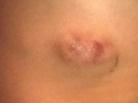
Anaplastic large cell lymphoma hidden by skin lesion
Non-Hodgkin's lymphomas are a group of diseases caused by neoplastic transformation of lymphoid cells. Anaplastic large cell lymphoma (<i>Anaplastic large cell lymphoma; </i><i>ALCL)</i>ALK+ (<i>anaplastic lymphoma kinase (ALK)-positive ALK+)</i>. is a neoplasm that originates from T lymphocytes. ALK is a tyrosine kinase receptor with an as yet undiscovered ligand so-called "orphan" (<i>orphan receptor</i>). Activation of of abnormal transmission is caused by a translocation involving the 2p23 region. ALK activation is thought to activates the STAT3, AKT/PI3K, RAS/ERK pathways responsible for cell proliferation and the cell cycle. The occurrence of ALCL ALK+ is relatively rare. It occurs in 3% of adult non-Hodgkin's lymphoma patients and in 10-15% of pediatric lymphoma patients. ALCL ALK+ is an aggressive lymphoma. It is mostly found in younger people (median age 34 years). It is more common in men than in women. The disease mainly occupies lymph nodes, but 60% of patients also have extranodal manifestations. The ALCL ALK+ staging classification uses the Ann Arbor classifications are used. Within 5 years of treatment, 60% of patients remain free of disease recurrence. [1]
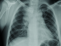
9 years old girl with pneumonia and congenital defect of pulmonary truct
Defects Congenital defects are abnormalities of the structure of organs created before birth, which lead to dysfunction of these organs. It is estimated that the formation of birth defects birth defects are influenced by: toxic-environmental factors (in 5-10%) and genetic factors (in 20-25%), but in 70-80% no cause can be determined. The incidence of all congenital malformations of congenital malformations in newborns and infants is about 4%, less than 4% of them (about 3.9%) involve the respiratory system. Nearly half of children with congenital defects of the respiratory system die before the age of five. Symptoms of defects in the respiratory system of the respiratory system include chronic or recurrent respiratory infections, symptoms of respiratory failure, coughing, and hemoptysis. Sometimes a defect in the respiratory system of the respiratory system is asymptomatic and detected accidentally.
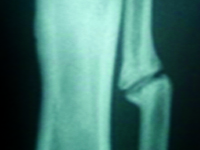
Pseudoarthrosis after tibia and fibula shaft fracture in a child – case report
Jest to rzadki przypadek, w którym pierwotnie zdiagnozowane przerzuty FTC do kości śródstopia nie zostały potwierdzone w dalszej diagnostyce. W związku z tym wykonano jedynie resekcję guza. Diagnostyka pooperacyjna opierała się na wielokrotnych, dokładnych badaniach histopatologicznych wycinków pobranych w trakcie operacji, co wykluczyło niekorzystne rozpoznanie. Dzięki temu uniknięto amputacji stopy.
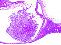
Acquired cystic disease associated renal cell carcinoma - new entity in classification WHO 2016. Report of two cases detected at an early stage of development.
W 2012 roku The International Society of Urological Pathology (ISUP) zaproponowało wdrożenie do klasyfikacji 5 nowych podtypów raka nerki, w tym raka nerki związanego z nabytą torbielowatością nerek (ang. acquired cystic disease - associated renal cell carcinoma - ACD-RCC). W 2016 roku ACD-RCC został uznany za niezależną jednostkę w Klasyfikacji Nowotworów Układu Moczowego WHO 2016. [1]. Poniższa praca prezentuje typowe cechy ACD-RCC w oparciu o dwa przypadki.
Diagnostic problems and therapeutic procedures in the case of neurosyphilis
Currently, syphilis is a growing epidemiological problem, as evidenced by the growing number of new infections, also in Poland [1]. A negative phenomenon is the increasing incidence of late-stage Treponema pallidum infections, including syphilis of the central nervous system (CNS) [2]. In advanced stages of syphilis, and especially in neurosyphilis, the risk of permanent complications increases, including tenesmus dorsi, progressive paralysis, stroke, and others [3]. CNS syphilis poses a significant diagnostic challenge because the symptoms of infection can mimic other primary CNS diseases or systemic diseases with secondary CNS involvement. Therefore, it is often called "the great imitator" [4, 5, 6, 7].
Foreign body of the neck - case report
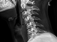
Foreign body ingestion is more common in children than in adults, but serious complications are less common in pediatric patients [1, 2]. Among adults, elderly individuals, those with masticatory disorders, esophageal strictures, prisoners, and patients with mental disorders predominate [2-4]. 80-90% of swallowed foreign bodies pass through the upper gastrointestinal tract spontaneously, but 10-20% require endoscopic removal, and 1% cause complications [3,5]. Possible locations of foreign bodies in the midpharynx include the epiglottis, the base of the tongue, the palatine tonsils, and in the hypopharynx, the piriform recesses, the retrocricoid region, and the posterior pharyngeal wall [6, 7]. In the esophagus, foreign bodies are located in the cervical part in over 80% of cases [1, 2, 8]. It is rare for a swallowed foreign body to penetrate the gastrointestinal tract and migrate deep into the soft tissues. This may be facilitated by the foreign body's structure, e.g., sharp-edged pieces of bone or glass. This poses a risk of serious and potentially fatal complications, such as retropharyngeal abscess, mediastinitis, or damage to the large vessels of the neck [7]. In such cases, prompt intervention and removal of the foreign body are essential. We present the case of a patient with a foreign body that migrated from the hypopharynx into the soft tissues of the neck, directly adjacent to the common carotid artery.