№ 43|Internal MedicineCase Report◯Open AccessFast review
Bilateral lung tumors - what to do?
Tumors were detected in a 59-year-old male long-term smoker with no significant past medical history
of both lungs.
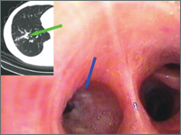
A bronchoscopy was performed, which showed bronchial stenosis on the right side to segment 9 by a
submucosal infiltration. Cytological examination of the brush biopsy smear and histopathological examination of the
sections from the lesion yielded a diagnosis of squamous cell carcinoma. Due to the equivocal
nature of the left lung lesion on CT scan, a PET-CT was performed. The result of the study:
SUVmax of right lung cancer = 1.9; left lung tumor = 8.8.It was decided to perform thoracotomy
left-sided thoracotomy. An ad hoc intraoperative cytological examination of the left lung tumor impression revealed
non-small cell carcinoma cells. The lower lobe was removed along with the mediastinal lymph nodes.
After two months, a video-guided right lower lobectomy with mediastinal lymphadenectomy was performed.
Histopathological examination of the right lung revealed pre-invasive squamous cell carcinoma
while the left lung was diagnosed as squamous cell carcinoma with an intermediate grade of cell differentiation
(G2). Taking into account the criteria of Martini and Melamed, bilateral synchronous foci of
of primary cancer.
Received 21 Jan 2013→Accepted 06 Feb 2013→Published 23 Mar 2013
BP
Błasiak P., Marciniak M., Pawełczyk K.23 Mar 2013